Don't worry, you don't need any if you start with the basics.
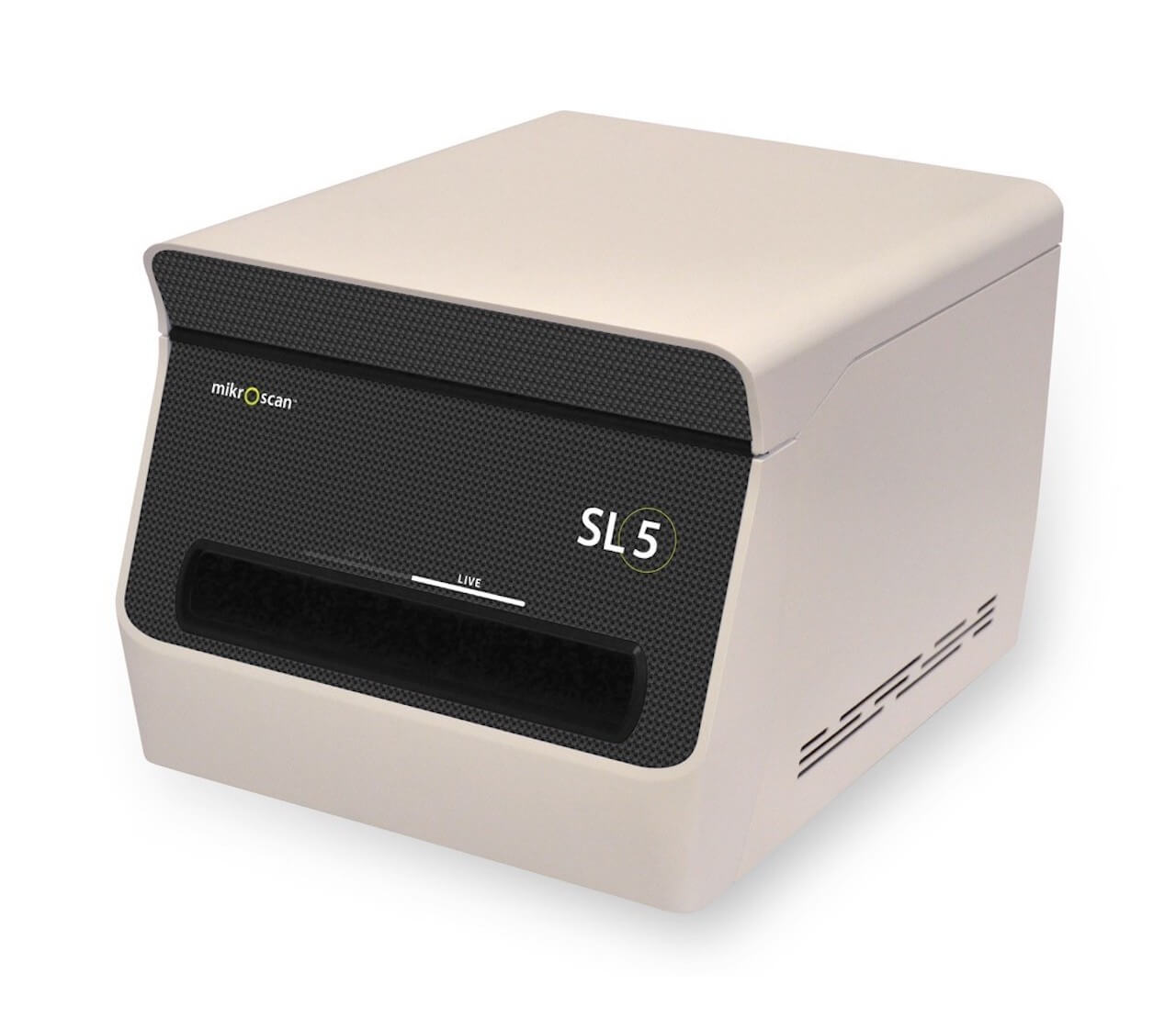
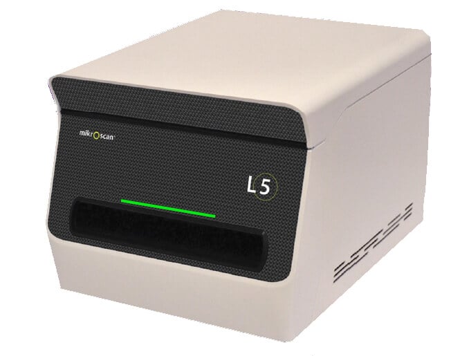
Telepathology that uses a Class I, remote robotic microscope to view slides LIVE, versus digitizing images with a scanner, will give you many of the benefits of digital pathology, but without the cost or time required to set it up.
With a telepathology system, you don't need to invest in desks, computers, staff, and training time.
Start with the basics, and build as your needs grow.
Learn more in our blog, On the Path:
Live telepathology, or telemicroscopy, distributes your pathology expertise across the hall, town and the world. Affordable, portable and simple to use solutions from Mikroscan help confirm adequate biopsies on the first pass, let you consult remotely in real-time, reduce travel, avoid unnecessary and expensive repeat procedures, and improve your bottom line.
It is imperative that we seek out solutions that help address the shift in reimbursement methodologies toward improved outcomes, reduced costs, and decreased risks. Mikroscan’s technology is a great tool for pathology departments to accomplish these objectives.
Keith Kaplan, MDPathologist |
We have a family of products to meet your pathology needs, from telemicroscopy systems to low- and medium-throughput digital pathology systems.
The SL5 Dual-Mode Real-Time Telemicroscopy and Digital Pathology System is the most compact, portable, and affordable telemicroscopy and digital pathology instrument on the market. It provides the added advantage of real-time telemicroscopy along with a completely separate mode for traditional "scan, store, and share" digital pathology applications. The SL5 contains a complete set of objectives—2X, 4X, 10X, 20X, and 40X—for true magnification of a wide range of applications and uses.
With the L5, you can be anywhere in the world and review a glass slide as though you were in the same room.
Using 5 high-quality microscope objectives, you will have complete control over slide navigation, focus, and illumination just like with a traditional brightfield microscope. You choose the planes of focus and areas of interest, not the system. Unlike with other systems, you can also control illumination from where you are to boost light for better examination of a thick specimen.
Trends in diagnostic interpretations reveal areas of improvement for current quality practices and provide vital opportunities for continuous improvement, patient safety, and positive outcomes. Read our case study to find out how to improve diagnostic proficiency in anatomic pathology.