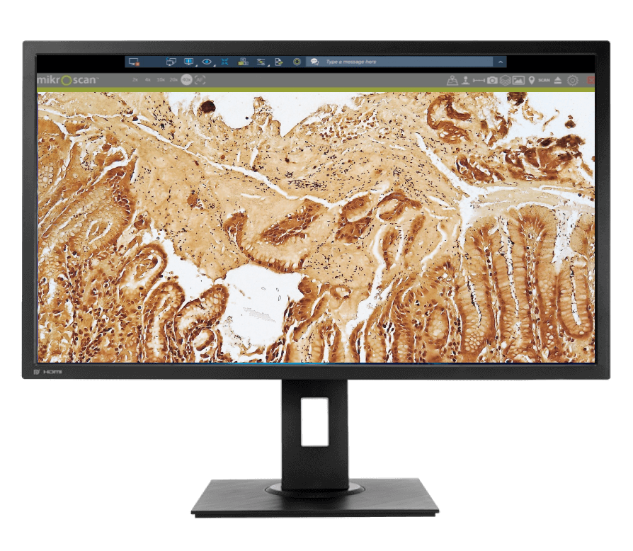
After a slide has been digitized through whole-slide scanning*, the digital image of the specimen can be analyzed by a variety of algorithms to automatically identify, segment, and categorize areas of interest for precise and highly reproducible results.
Research laboratories, such as those for pharmaceutical and academic research, performing tissue-based research were the earliest adopters of image analysis due to the technology's ability to score biomarker expression quickly and objectively.
The use of image analysis routines has been demonstrated to improve reproducibility and accuracy in quantitative applications. The next generation of decision support tools, such as machine learning, will provide deeper analysis and insights not available through current techniques.
Our Solutions
The non-proprietary TIF format of our SL5 and SL5-20 whole-slide scanners allows you to perform a variety of image analysis applications.
Our image analysis-ready digital slides allow for quantitative analysis such as membrane, nuclear and cytoplasm biomarker quantification. Tissue classification, morphology, and other imaging applications like brightfield-based in situ hybridization (ISH) are also possible.*
Current image analysis solutions are provided by HALO by Indica Labs, Inc., and Visiopharm®