Tumor boards are meetings where specialists from surgery, medical oncology, radiation oncology, radiology, genetics, and pathology collaboratively review a patient's condition and determine the best treatment plan.
With so many multi-disciplinary teams, the ability to remotely share and collaborate on pathology images is critical.
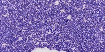
With live telemicroscopy, pathology images used in tumor boards now come alive, augmenting other visuals and data residing in the patient’s electronic medical record. Pathologists can demonstrate findings in real-time via interactive images, and address questions and interpretations with tumor board colleagues using the remote, robotic microscope.
Pathologist peer review or working groups are no longer constrained to discussions in a single location and a multi-headed microscope, and all participants benefit from an interactive review with the pathologist.
We provide the L5 system, a Class I, remote robotic microscope dedicated to telemicroscopy. Our SL5 system is dual-mode, and includes the robotic microscope of the L5 for live telemicroscopy applications and a whole-slide scanner for digital pathology* applications.

Live telemicroscopy distributes your pathology expertise across the hall, town, and the world. Affordable, portable, and simple-to-use solutions from Mikroscan help confirm adequate biopsies on the first pass, let you consult remotely in real-time, reduce travel, avoid unnecessary and expensive repeat procedures, and improve your bottom line.