
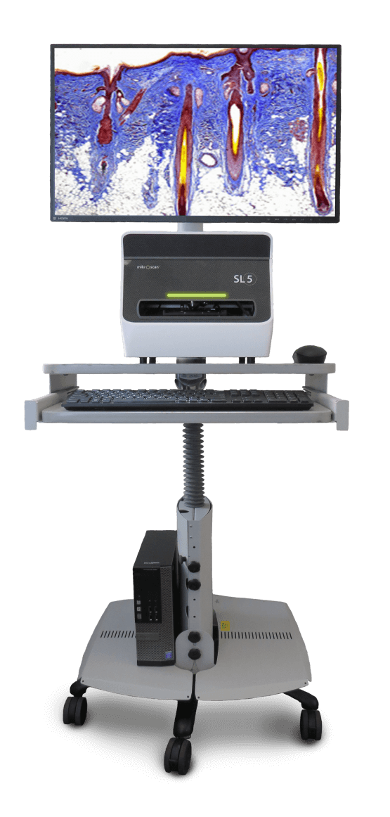
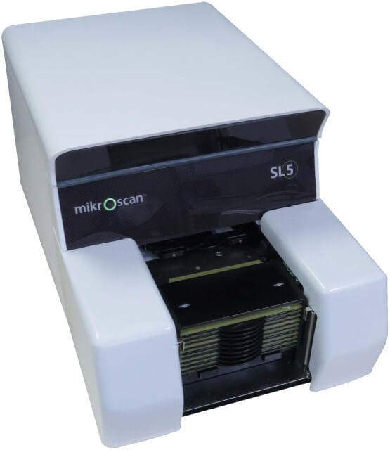
The only system that operates as a live robotic microscope or digital pathology scanner.
Our dual-mode system offers two completely separate and distinctly different capabilities in one convenient device. You select one of two modes—a live robotic microscope for the examination of specimens on glass slides or a static digital pathology scanning mode for the creation of digitized tissue samples.*
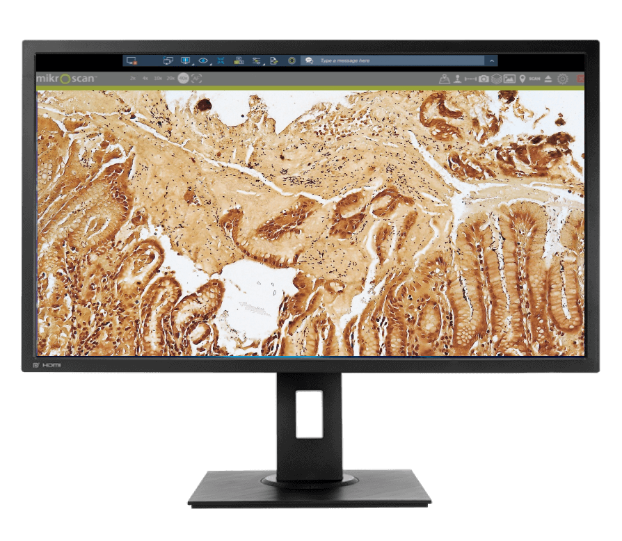
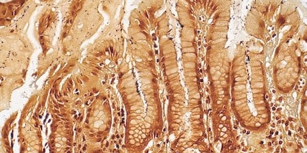
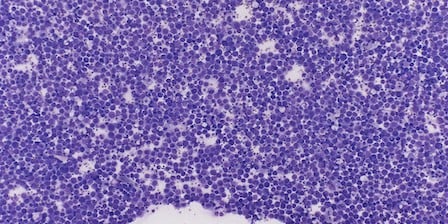
In Scan Mode, create whole-slide images for research applications that may require image analysis, educational applications such as tumor boards, quality assurance, or for storage and archival purposes.
Live Mode uses our live robotic microscope to perform remote examinations for clinical applications such as ROSE, frozen sections, and subspecialty consultations.

I have used 3-4 other digital pathology scanners and this is by far the best product I have used for frozen section consultation.
Henry O’DellChief, Clinical Operations, Keck School of Medicine, USC |


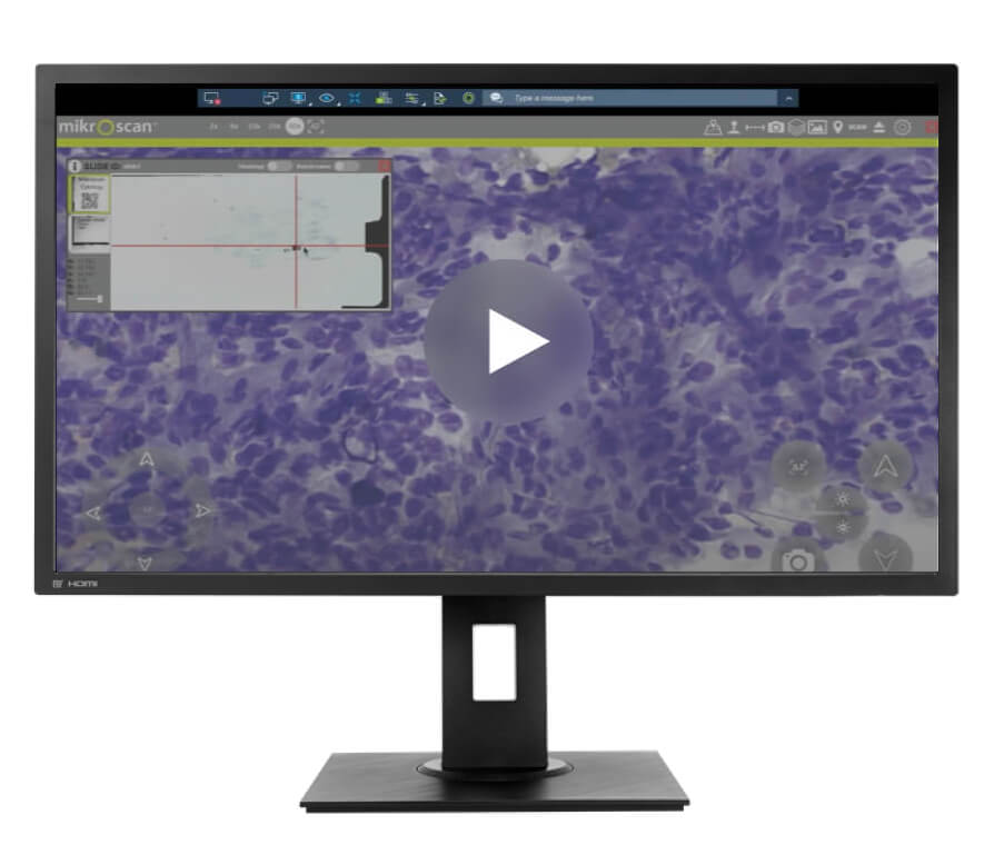
See how the Live mode of the SL5 system allows pathologists to evaluate glass slides from anywhere in the world.
We have a family of products to meet your pathology needs, including telemicroscopy systems and low- and medium-throughput digital pathology systems.
Rapidly review entire cases with our new, 20-slide capacity L5-20 Real-Time Telemicroscopy and SL5-20 Dual-Mode Real-Time Telemicroscopy and Digital Pathology systems.* The 20-slide capacity mirrors traditional pathology folders, and with just a 33% increase in bench depth, the 5-20 systems have the smallest footprint of any system at this performance level.


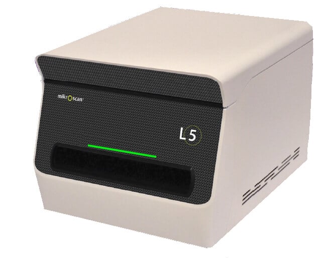
With the L5, you can be anywhere in the world and review a glass slide as though you were in the same room.
Using 5 high-quality microscope objectives, you will have complete control over slide navigation, focus, and illumination just like with a traditional brightfield microscope. You choose the planes of focus and areas of interest, not the system. Unlike with other systems, you can also control illumination from where you are to boost light for better examination of a thick specimen.


Download the Technical Specs for our SL5 system to learn about system information and requirements.
