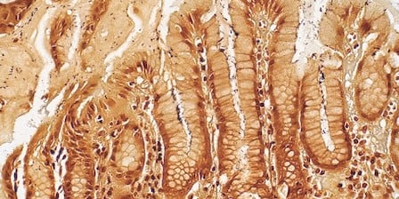
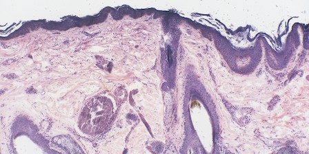
Disease research in pathology informatics requires archiving, retrieving, organizing, sharing, and analyzing diverse pathology-related data sources. The use of digital pathology* to create whole-slide images provides the opportunity for advanced archival and retrieval capabilities.
Laboratories implementing a digital workflow can securely store and retrieve whole-slide images along with associated case meta-data. This convenience helps to organise archival systems better and eliminates concern over breakage, loss of glass slides, and degradation over time.
Sharing and Image Analysis
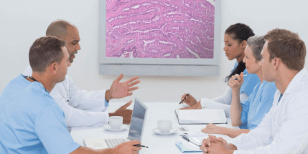
The archival and easy retrieval of pathology images also facilitates sharing and analysing data across different groups. Today's research environment involves multiple interdisciplinary teams located around the world who can easily use a digital archive to share images with each other and then collaborate in the laboratory or cloud.¹
Our Solutions
We provide the low-throughput, 2-slide capacity SL5 system and the medium-throughput, 20-slide capacity 5-20 system for research, quality assurance, and education.